Quick Answer: An ultrasound scan uses high-frequency sound waves — not X-rays or radiation — to create real-time images of your body’s internal organs and structures. It is safe, painless, and suitable for all ages, including pregnant women and children. If you are experiencing unexplained symptoms or want a preventive health check, Best Ultrasound Clinic in Brentwood offers private ultrasound scans across Essex with same-day appointments and no GP referral book online.
Ultrasound scans are one of the most common and versatile medical imaging tests in use today. Yet many patients arrive at their scan appointment unsure of what to expect, how the procedure works, or whether they actually need one. This easy-read guide is designed to help you understand the world of diagnostics — and specifically, when and why an ultrasound scan might be exactly what your health requires.
Whether you have been advised by a doctor, noticed a new symptom, or simply want to learn more about your health, this guide will walk you through everything you need to know — from the basics of how sound waves create images, to the different types of scans available at our private clinic in Brentwood, Essex.
Table of Contents
Ultrasound Education: What Is an Ultrasound Scan and How Does It Work?
Ultrasound imaging — also called sonography — is a medical examination that uses high-frequency sound waves to produce detailed pictures of structures inside the body. Unlike a CT scan or standard X-rays, ultrasound does not use ionising radiation, making it one of the safest diagnostic tools available to medical providers worldwide.
The process is straightforward: a small handheld device called a probe (or transducer) is placed against the skin, which has been coated with a water-based gel. The gel ensures smooth contact and helps sound waves travel efficiently into the body. The probe emits high-frequency sound waves — typically in the range of 2 to 18 megahertz — which travel through soft tissue and bounce back when they encounter different structures or organs.
These returning echoes are captured by the probe and processed by specialised equipment, which converts the sound data into a real-time image displayed on a screen. This image is called a sonogram. Depending on the density and composition of the tissue, different structures appear in varying shades of grey — from bright white (hyperechoic, such as bone surfaces) to dark black (anechoic, such as fluid-filled structures like the bladder).
Understanding What You See on Screen
- Homogeneous appearance: Tissue that looks uniform in texture and echogenicity — a sign of normal, healthy organ structure.
- Heterogeneous appearance: Irregular, uneven texture within an organ — which may indicate inflammation, cysts, fibroids, or tumours requiring further diagnosis.
- Anechoic (black): No echo returned — usually fluid such as urine in the bladder, amniotic fluid, or blood vessels.
- Hyperechoic (bright white): Strong echo — bone surfaces, gallstones, or calcified tissue.
Your radiologist or trained sonographer will interpret these appearances in context with your clinical history to produce a clear, accurate diagnosis or reassurance of normal findings.
Sound Waves, Not Radiation: Is an Ultrasound Scan Safe?
One of the most reassuring facts about ultrasound imaging is that it is entirely safe for patients of all ages. Because the procedure relies solely on sound waves — not X-rays, CT radiation, or magnetic fields — it carries no known risks when performed by a trained specialist using appropriate equipment.
Ultrasound scans are safe to perform during pregnancy (including on a baby at any stage), on newborns and children, on elderly patients, and on individuals with metal implants or pacemakers who may be unsuitable for MRI. There is no cumulative dose, no recovery time, and no need for sedation or fasting in most cases.
| Feature | Ultrasound | CT Scan | MRI |
| Radiation | None — sound waves only | Yes (ionising X-rays) | None — magnetic field |
| Safe in Pregnancy | Yes | No (avoid if possible) | Usually yes |
| Cost | Low–moderate | Moderate | High |
| Availability | Widely available | Hospital-based | Specialist centres |
| Real-Time View | Yes — dynamic imaging | No | No |
| Soft Tissue | Excellent | Good | Excellent |
| Bone Detail | Limited | Excellent | Good |
| Guide Injections | Yes | Sometimes | Rarely |
For most soft tissue conditions — including abdominal pain, pelvic symptoms, joint and tendon injuries, and pregnancy monitoring — ultrasound scans provide excellent image quality, real-time visualisation, and a comfortable examination experience. CT and MRI remain essential tools in specific clinical scenarios, but for the broad range of conditions evaluated in primary and specialist care, ultrasound is frequently the first-choice imaging modality.
Types of Ultrasound Scans
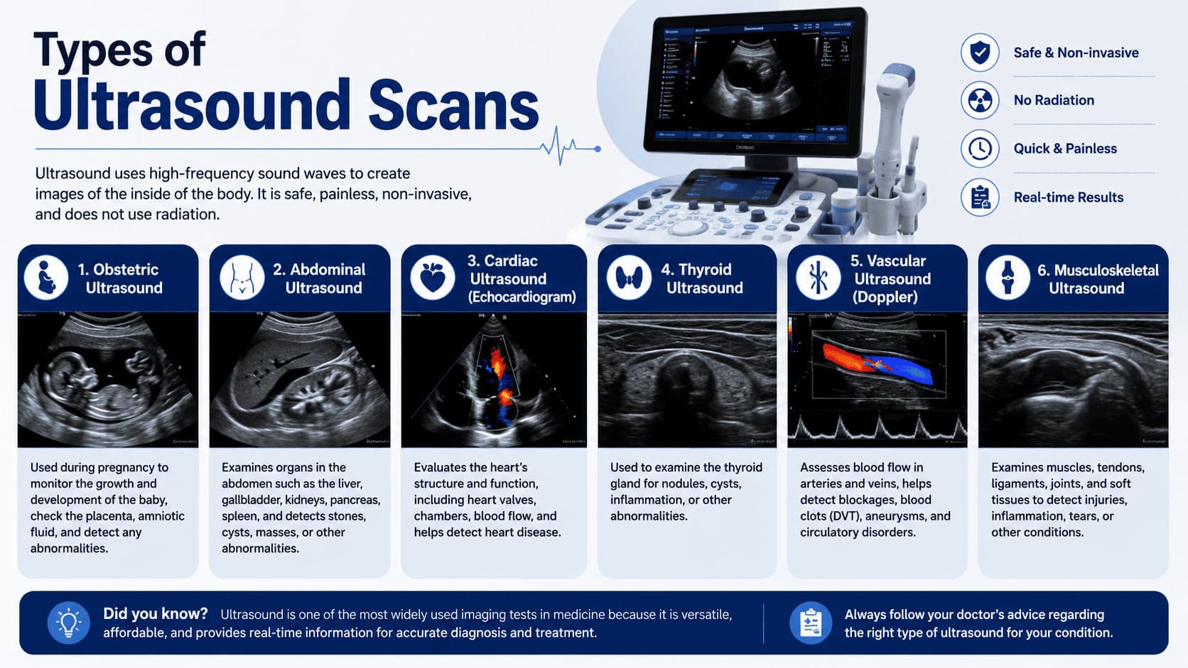
Entering the world of diagnostics can feel overwhelming, particularly when a medical provider mentions a type of scan you have never heard of. Here is a clear overview of the most common imaging tests performed using ultrasound technology, and the medical conditions each one is designed to evaluate.
| Scan Type | Best Used For | Who Performs It |
| Abdominal | Liver, gallbladder, kidneys, spleen, pancreas | Sonographer / Radiologist |
| Pelvic | Uterus, ovaries, bladder (trans-abdominal) | Sonographer / Radiologist |
| Transvaginal | Detailed uterine/ovarian imaging via internal probe | Specialist Sonographer |
| Pregnancy Baby | Fetal development, heartbeat, growth, anatomy | Obstetric Sonographer |
| Thyroid | Thyroid nodules, goitre, lymph nodes | Radiologist |
| Venous Doppler | DVT, blood clots, varicose veins, blood flow | Vascular Radiologist |
| Musculoskeletal | Tendons, ligaments, muscles, joints, bursae | MSK Radiologist |
| Whole Body | Preventive health screening, multiple organs | Consultant Radiologist |
| 3D / 4D Baby | Bonding scan, baby’s facial features, real-time movement | Obstetric Sonographer |
Pelvic Ultrasound for Women and Children
Pelvic ultrasound is one of the most commonly requested ultrasound scans for women and is performed routinely for a wide range of gynaecological and obstetric indications. The pelvic examination uses high-frequency sound waves to create detailed images of the uterus, ovaries, fallopian tubes, and bladder — structures that are difficult to assess without imaging.
For women experiencing irregular or heavy periods, unexplained pelvic pain, suspected fibroids, ovarian cysts, or symptoms of endometriosis, a pelvic ultrasound scan is typically the first imaging step. For children and young patients, the procedure is non-invasive and entirely painless, making it a safe and practical diagnostic option.
Where greater detail is required — for example when assessing early pregnancy, evaluating endometrial thickness, or investigating suspected polyps — a transvaginal ultrasound may be recommended. This uses a small, smooth probe gently placed internally to produce higher-resolution images of the pelvic organs. The procedure is straightforward and typically takes under ten minutes when performed by an experienced specialist.
Venous Doppler and Blood Flow Studies
A venous Doppler scan is a specialist type of ultrasound examination that assesses blood flow within the veins and arteries. Using colour Doppler technology, the scan can detect deep vein thrombosis (DVT), varicose vein reflux, carotid artery narrowing, and peripheral arterial disease. Blood appears as a moving, colourful signal on the image, making it easy for the specialist to identify where flow is normal, reduced, or blocked.
Venous Doppler studies are especially important for patients who have experienced leg swelling, pain, or skin changes following prolonged immobility, surgery, or long-haul travel. Results are provided step by step during the examination, with a full written report available the same day.
When Should You Book an Ultrasound Scan Appointment?
Ultrasound scans are appropriate across a wide range of clinical and preventive health scenarios. The following are the most common reasons patients at our Essex clinic book a scan appointment — with or without a GP’s advice.
Symptoms That Warrant an Ultrasound Scan
- Abdominal pain or discomfort: Unexplained pain in the abdomen may indicate gallstones, kidney stones, liver disease, or pancreatic conditions — all visible on a standard abdominal scan.
- Pelvic pain or pressure: Pain in the lower abdomen or pelvis in women may point to fibroids, ovarian cysts, endometriosis, or pelvic inflammatory disease.
- Bloating or a feeling of fullness: Persistent bloating can be a sign of organ enlargement, fluid accumulation, or structural changes detectable on an ultrasound scan.
- A lump or swelling: Any new lump — whether in the neck, abdomen, breast area, or soft tissue — should be assessed with a targeted ultrasound to guide a clear diagnosis.
- Urinary symptoms: Difficulty urinating, blood in urine, or frequent urges may be linked to bladder wall changes, kidney abnormalities, or prostate enlargement.
- Joint or tendon pain: Musculoskeletal ultrasound scans can identify rotator cuff tears, Achilles tendinopathy, ligament injuries, and bursitis with high accuracy.
- Leg pain or swelling: Particularly after inactivity, surgery, or travel — an urgent DVT scan appointment may be needed to rule out a blood clot.
Preventive and Monitoring Scans
- Annual wellbeing check for men and women over 40
- Thyroid monitoring for patients with known nodules or abnormal thyroid function tests
- Abdominal aortic aneurysm screening for men over 65 with cardiovascular risk factors
- Liver health monitoring for patients with fatty liver disease or alcohol history
- Ovarian and uterine monitoring for patients with PCOS or fibroid history
Pregnancy-Related Scans
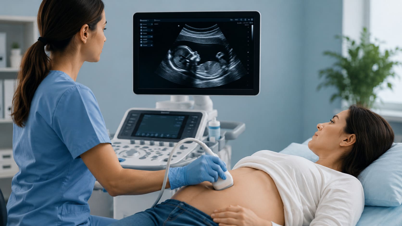
- Early pregnancy scan (6–12 weeks) to confirm heartbeat, dating, and location
- Nuchal translucency scan (11–14 weeks) for chromosomal risk assessment
- 20-week anatomy scan — the most detailed pregnancy examination
- Growth and Doppler scans in the third trimester for high-risk pregnancies
- 3D/4D baby scan for bonding and detailed facial imaging (26–32 weeks)
Private Ultrasound Scan Essex
For patients across Essex who need fast, expert imaging without NHS waiting times, Best Ultrasound Clinic provides a full range of private ultrasound scans from our Brentwood clinic. Whether your scan appointment is for a specific medical concern or a routine preventive check, you will receive the same high standard of care: state-of-the-art ultrasound equipment, a trained consultant radiologist, and a same-day written report.
Private ultrasound scans in Essex are accessible without a GP referral — you can book directly by phone, online, or via WhatsApp. Appointments are available Monday to Friday from 8am to 8pm and on Saturdays, making it easy to fit your health into a busy schedule.
What Is Included in Every Private Scan Appointment?
Consultation with a specialist radiologist or trained sonographer — real-time imaging using the latest ultrasound equipment — clear explanation of findings during the examination — same-day written report and images — referral letter if follow-up care is required — no hidden charges or unexpected fees.
Conditions We Diagnose at Our Essex Clinic
- Gallstones, kidney stones, and liver disease
- Ovarian cysts, fibroids, and endometrial abnormalities
- Thyroid nodules, goitre, and lymph node enlargement
- Deep vein thrombosis and venous insufficiency
- Musculoskeletal injuries including tendon and ligament tears
- Pregnancy complications and fetal growth monitoring
- Testicular abnormalities and scrotal conditions
- Lumps, bumps, and soft tissue masses anywhere on the body
Ultrasound Scan Near Me Brentwood
If you have searched for an ultrasound scan near me and you are based in Brentwood or the surrounding Essex area, Best Ultrasound Clinic is your nearest specialist private imaging centre. Located at 27 Kings Road, Brentwood, CM14 4DJ, our clinic is easily accessible by car and public transport from across Essex.
We provide imaging services for patients from Brentwood, Chelmsford, Colchester, Basildon, Romford, Billericay, and beyond. Our consultant radiologists are specialists in musculoskeletal, obstetric, vascular, and abdominal ultrasound — covering the full clinical range within a single clinic.
How to Book Your Scan Appointment
- Online: Visit bestultrasoundclinic.co.uk and use the Book Now button — available 24/7
No referral is ever needed. Simply choose your scan type, select a convenient time, and attend your appointment. Your specialist will guide you through every step of the procedure and make sure you leave with a clear picture of your health.
NHS vs. Private Ultrasound: Which Is Right for You?
The NHS provides excellent ultrasound services and remains the right choice for many patients. However, long waiting times — often six to eighteen weeks for non-urgent scans — mean that patients with worsening symptoms, anxiety, or time-sensitive conditions may benefit significantly from accessing private ultrasound care.
The following comparison helps users understand the practical differences between NHS and private ultrasound services in Essex.
| Criteria | NHS Ultrasound | Private Ultrasound Essex |
| Waiting Time | 6–18 weeks | Same day or within days |
| Referral Required | Yes — GP/consultant referral | No — self-refer directly |
| Appointment Slots | Weekdays only | Evenings & weekends available |
| Scan Duration | 15–20 min, often rushed | 30–45 min, unhurried |
| Results | Posted after days/weeks | Same-day verbal & written report |
| Choice of Scan | GP decides which scan | You choose or specialist advises |
| Follow-Up | Return to GP then re-refer | Specialist consultation included |
| Cost | Free at point of use | Transparent private pricing |
Many patients choose to use private ultrasound for initial diagnosis and peace of mind, then share the report with their GP to guide further NHS treatment if required. Others use private scans to monitor known conditions between NHS appointments, or to access specialist imaging — such as musculoskeletal or vascular studies — that may not be readily available at their local NHS trust.
Is a Private Ultrasound Report Accepted by NHS Doctors?
Yes. A report from a qualified consultant radiologist at a private clinic carries the same clinical weight as an NHS report. Your GP or hospital specialist can use the findings to guide your care, request further investigations, or initiate treatment. We always provide a professional written report with images that you are free to share with any medical provider.
What to Expect at Your Scan Appointment: Step by Step
For patients attending their first ultrasound scan — or returning after a long break — here is a simple, step-by-step guide to what happens during a typical appointment at our Brentwood clinic.
Step 1 — Preparation
Preparation depends on the type of scan. For abdominal scans, you will be asked to fast for six hours before your appointment so that the stomach and bowel do not obstruct the view of deep organs. For pelvic scans, you will need a full bladder — drink one litre of water one hour before and do not empty it. For most other scans, including thyroid, musculoskeletal, and pregnancy ultrasound, no preparation is needed at all.
Step 2 — Arrival and Consultation
You will be welcomed by our team and asked to complete a brief health questionnaire. Your specialist will then discuss your symptoms and medical history to ensure the scan is focused on the most clinically relevant areas. This is your opportunity to ask any questions and raise any concerns.
Step 3 — The Examination
You will lie on a comfortable examination couch. A small amount of warm water-based gel is applied to the skin over the area being scanned. The sonographer or radiologist places the probe gently on the skin and moves it in a systematic way to produce images of the organs and structures being examined. The examination is entirely painless. You may be asked to hold your breath briefly, change position, or point to where you feel any discomfort.
Some examinations — such as transvaginal pelvic scans or internal prostate assessments — require an internal probe. Your specialist will explain exactly what is involved beforehand, and you are always free to ask questions or decline any aspect of the procedure.
Step 4 — Results and Report
One of the key advantages of private ultrasound is that your doctor or radiologist will explain the scan findings to you directly — in plain language — before you leave. You will receive a written report on the same day, along with printed images where appropriate. If any follow-up care, further imaging, or specialist referral is recommended, our team will guide you through the next steps clearly and without delay.
Book Your Private Ultrasound Scan in Essex Today
Whether you have a specific symptom to investigate, a condition to monitor, or simply want the peace of mind that comes from a thorough health check, our team at Best Ultrasound Clinic is here to help. We offer the full range of ultrasound scans — from abdominal and pelvic examinations to specialist vascular, musculoskeletal, and obstetric imaging — all performed by qualified consultant radiologists in a comfortable, welcoming environment.
Best Ultrasound Clinic | 27 Kings Rd, Brentwood, Essex CM14 4DJ
📧 info@essexprivateclinicultrasound.co.uk | 🌐 bestultrasoundclinic.co.uk
Frequently Asked Questions — Ultrasound Scans Essex
Does an ultrasound scan hurt?
No. Ultrasound is a completely painless examination. You may feel a little pressure from the probe, and the gel can feel slightly cool, but there is no discomfort during a standard external scan. Internal scans (such as transvaginal) use a small smooth probe and are performed gently by a trained specialist.
How long does an ultrasound scan take?
Most standard ultrasound scans take between 15 and 30 minutes. More detailed examinations — such as a whole-body wellbeing scan or a detailed 20-week pregnancy scan — may take 30 to 45 minutes. You will always be told how long to expect at the time of booking.
Do I need a doctor’s referral to book a private ultrasound scan in Essex?
No. Best Ultrasound Clinic accepts direct bookings from patients — no GP or specialist referral is required. You can book your scan appointment online, by phone, or via WhatsApp at any time.
Can I use a private ultrasound report with my NHS GP?
Yes. A report from our consultant radiologist is a recognised clinical document that your GP or hospital specialist can use to guide your further care. Many patients share their private scan results with their NHS team to speed up diagnosis and treatment.
What is the difference between a sonographer and a radiologist?
A sonographer is a trained medical professional who specialises in performing and interpreting ultrasound scans. A radiologist is a qualified doctor who has undergone additional specialist training in medical imaging, including ultrasound, CT, and MRI. At Best Ultrasound Clinic, all scans are overseen by a consultant radiologist to ensure the highest standard of diagnosis.
Are ultrasound scans safe for children?
Yes, ultrasound scans are safe for patients of all ages, including newborns and children. They use sound waves rather than radiation, making them the preferred imaging choice for paediatric patients.
Can ultrasound detect cancer?
Ultrasound can identify suspicious masses, lymph node changes, and tissue abnormalities that may warrant further investigation — including biopsy or additional imaging such as MRI or CT. Ultrasound alone does not diagnose cancer, but it plays a vital role in early detection and guiding specialist care.
How is a pelvic ultrasound different from a transvaginal scan?
A pelvic ultrasound scan is performed externally — the probe is placed on the lower abdomen and requires a full bladder to produce clear images. A transvaginal scan uses a small internal probe to image the uterus and ovaries from closer range, providing higher resolution pictures — particularly useful in early pregnancy and detailed gynaecological assessment.