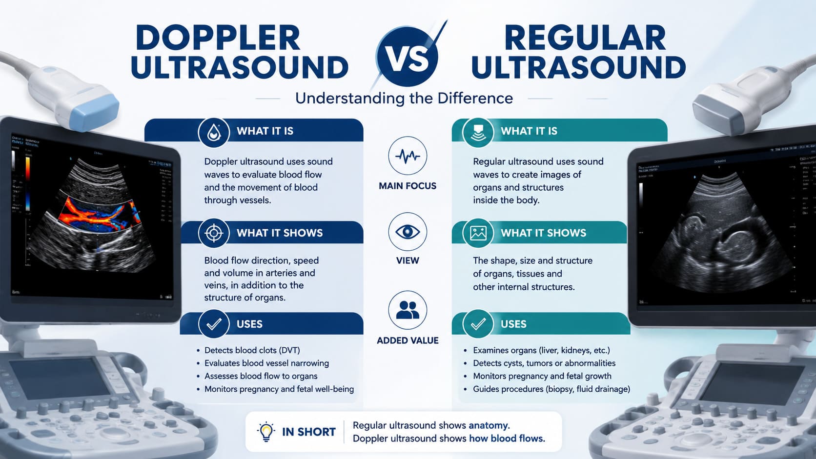
Quick Answer: A regular ultrasound uses high-frequency sound waves to produce structural images of organs and tissues. A Doppler ultrasound adds a second layer of technology — it measures the speed and direction of blood flow through veins and arteries, displaying this information in colour on screen. If you need a venous Doppler scan, carotid Doppler, or urgent DVT scan in Essex, Best Ultrasound Clinic in Brentwood offers same-day private appointments with no GP referral.
If you have been told you need a Doppler test and are wondering how it differs from the plain ultrasound you may have had before, you are not alone. The terms are often used interchangeably in everyday conversation — but in the world of diagnostics, they describe two distinct imaging techniques with very different clinical applications.
Understanding whether you need a regular ultrasound or a Doppler ultrasound — and why — can help you ask better questions at your scan appointment, choose the right type of examination, and make sense of your results. This article explains doppler ultrasound vs regular ultrasound clearly, compares their applications, and guides you through the Doppler services available at our private clinic in Brentwood, Essex.
Table of Contents
What Is Regular Ultrasound? Understanding Plain Ultrasound Today
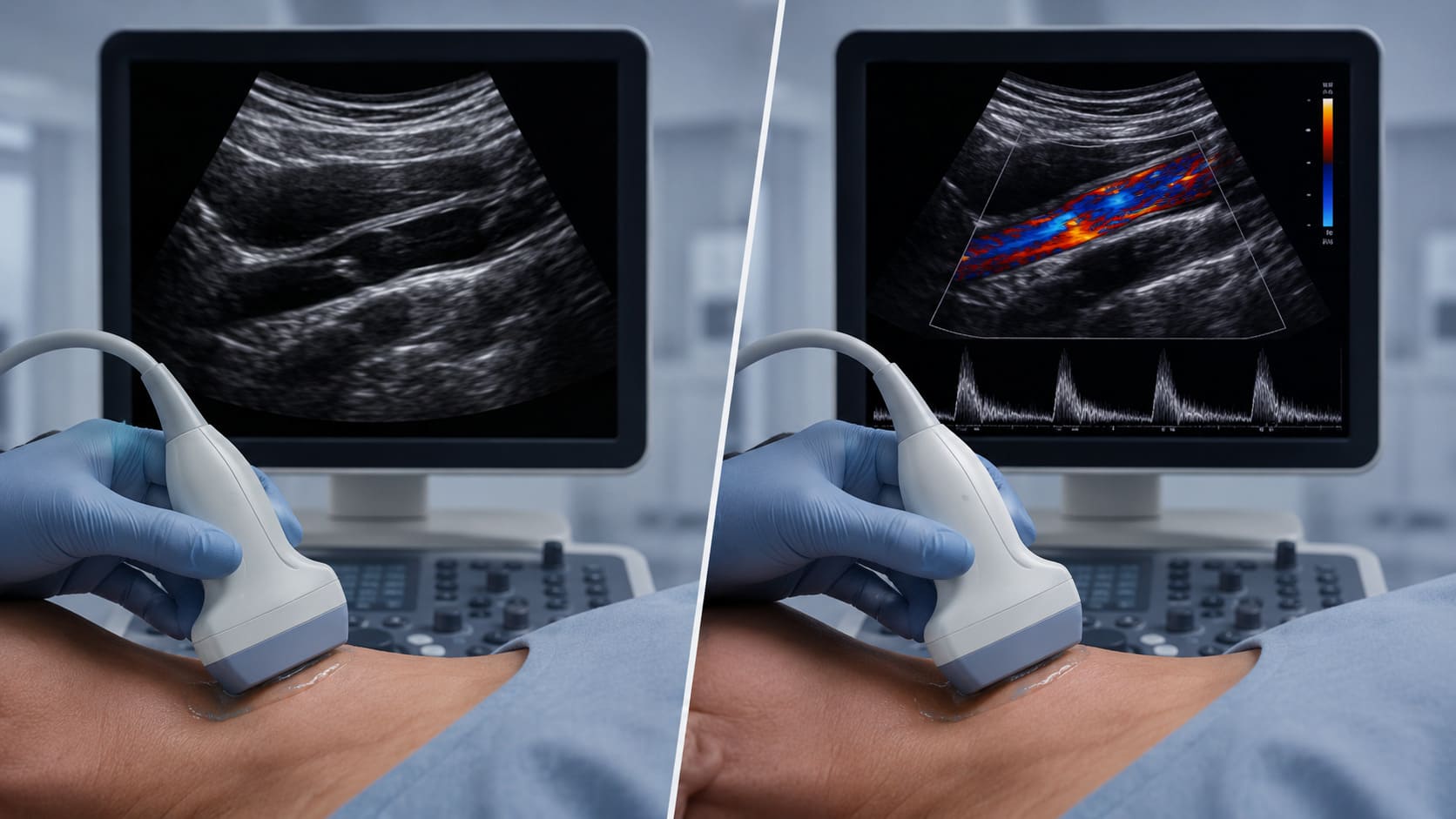
Regular ultrasound — also called plain ultrasound, standard ultrasound, or B-mode (brightness mode) ultrasound — is the most widely used imaging technique in clinical practice today. It produces real-time, two-dimensional images of the body’s internal anatomical structures by emitting high-frequency sound waves from a handheld probe and capturing the returning echoes as they bounce off different tissues.
These echoes are processed by the ultrasound equipment to create a greyscale image — a visual map of the structural landscape inside your body. Organs appear in different shades depending on their density: fluid appears black (anechoic), solid organs such as the liver appear mid-grey, and highly reflective surfaces such as bone appear bright white.
What Regular Ultrasound Can Show
- Anatomical structures: Size, shape, position, and internal texture of organs — liver, kidneys, gallbladder, spleen, pancreas, thyroid, uterus, ovaries, testes.
- Structural abnormalities: Cysts, masses, fibroids, nodules, gallstones, kidney stones, and organ enlargement.
- Soft tissue injuries: Tendon tears, ligament damage, muscle injuries, and joint effusions visible on musculoskeletal scans.
- Pregnancy monitoring: Fetal development, heartbeat, position, placenta location, and amniotic fluid levels.
- Diagnostic evaluation: Assessment of lumps, bumps, and swellings anywhere in the body.
Regular ultrasound is the starting point for most diagnostic imaging pathways. It is non-invasive, cost-effective, fast, and available without radiation. However, one critical limitation is that plain ultrasound cannot directly assess blood flow — it can only show structural parameters. This is where the addition of Doppler technology transforms ultrasound into a vascular diagnostic tool.
What Is Doppler Ultrasound?
Doppler ultrasound is a specialised extension of regular ultrasound that uses the Doppler effect — a fundamental principle of physics — to measure the movement of blood through vessels in real time. Named after Austrian physicist Christian Doppler, the technique detects changes in the frequency of sound waves as they reflect off moving red blood cells. When blood flows towards the probe, the returning frequency is higher; when blood flows away, the frequency is lower.
These frequency differences are converted by the ultrasound equipment into visual and audible information: blood flow speed, direction, volume, and resistance are all displayed on screen — typically as a colour overlay on top of the standard greyscale structural image. This is what is known as colour Doppler.
The Doppler Effect in Clinical Practice
The addition of Doppler technology transforms ultrasound from a static picture of anatomy into a dynamic evaluation of circulation. Instead of simply showing that a vein exists, a Doppler test can confirm whether blood is flowing through it normally, sluggishly, or not at all — a distinction that is critical for diagnosing conditions such as deep vein thrombosis (DVT), carotid artery stenosis, and renal artery hypertension.
How Colour Doppler Works
In colour Doppler imaging, blood flowing towards the probe is displayed in red, while blood flowing away is displayed in blue. The intensity of the colour indicates flow speed — brighter colours represent faster flow. Areas of absent colour signal indicate no flow, which may suggest a clot, occlusion, or significant narrowing. Spectral Doppler adds a waveform graph below the image, allowing the radiologist to measure precise velocity parameters and resistance indices for clinical evaluation.
Compare: doppler ultrasound vs regular ultrasound — Key Differences
The table below provides a clear, side-by-side comparison of plain ultrasound and Doppler ultrasound across the most important diagnostic parameters. Use this to discover which type of scan is most likely to be recommended for your specific clinical situation.
| Feature | Regular Ultrasound (Plain) | Doppler Ultrasound |
| Primary Function | Images anatomical structures & organs | Images structures + measures blood flow |
| Technology Used | High-frequency sound waves (echo) | Sound waves + Doppler frequency shift analysis |
| What It Shows | Size, shape, texture, structural appearance | Blood flow direction, speed, volume, resistance |
| Colour Display | Greyscale (black, white, grey) | Colour overlay (red/blue for flow direction) |
| Best For | Organs, cysts, masses, pregnancy, tendons | Veins, arteries, DVT, varicose veins, carotid |
| Detects Clots | Limited — structural changes only | Yes — directly visualises absent/reduced flow |
| Real-Time Flow | No | Yes — dynamic, moment-by-moment monitoring |
| Clinical Use | Diagnosis, screening, monitoring | Vascular evaluation, surgical planning, DVT |
| Exam Duration | 15–25 minutes | 20–40 minutes depending on vessels assessed |
| Radiation | None | None |
In practice, most Doppler examinations begin with a standard regular ultrasound to assess the structural appearance of the vessels and surrounding tissues, before colour Doppler is applied to evaluate blood flow. The two techniques therefore complement rather than compete with each other — Doppler is an addition to, not a replacement for, plain ultrasound imaging.
Types of Doppler Ultrasound: Which One Do You Need?
Doppler ultrasound is not a single, fixed technique. Modern vascular imaging uses several different Doppler modes, each with distinct applications and diagnostic strengths. Here is a practical overview of the techniques used at our Essex clinic, and the conditions each one is best suited to evaluate.
| Doppler Type | How It Works | Clinical Application |
| Colour Doppler | Colour codes flow direction: red = towards probe, blue = away | DVT, carotid, renal vessels, portal vein |
| Power Doppler | Detects very slow or low-volume flow using signal amplitude | Tumour vascularity, testicular blood flow |
| Spectral (Pulsed) | Produces waveform graph showing flow velocity over time | Carotid stenosis, renal artery, obstetric Doppler |
| Continuous Wave | Constantly emits and receives signal for fast, high-velocity flow | Cardiac valve assessment, arterial stenosis |
| 3D / 4D Doppler | Three-dimensional colour flow reconstruction in real time | Fetal circulation, complex vascular anatomy |
For the majority of patients attending a private clinic for a venous Doppler scan or DVT assessment, colour Doppler combined with spectral analysis is the standard approach. For carotid artery evaluation, spectral Doppler is essential to measure velocity parameters and calculate stenosis grade. Your specialist will select the most appropriate Doppler technique based on your clinical presentation and referral information.
Clinical Applications: When Do You Need a Doppler Test?
The following conditions and clinical scenarios represent the most common reasons patients are referred for a Doppler ultrasound. In each case, standard plain ultrasound would be insufficient on its own — blood flow evaluation is critical to reach an accurate diagnosis.
Deep Vein Thrombosis (DVT) and Pulmonary Embolism
DVT is one of the most time-sensitive diagnoses in medicine. A blood clot in the deep veins of the leg — if not identified and treated promptly — can travel to the lungs as a pulmonary embolism, a potentially life-threatening emergency. Doppler ultrasound is the first-line diagnostic test for DVT. By directly visualising blood flow (or its absence) in the deep leg veins, a Doppler test can confirm or exclude a clot within minutes.
Regular ultrasound alone cannot reliably detect a DVT — it can identify a thickened vein wall or subtle structural changes, but it cannot assess flow. This makes Doppler technology critical for any patient with leg pain, swelling, redness, or warmth following surgery, long-haul travel, or prolonged immobility.
Varicose Vein Assessment and Venous Insufficiency
Varicose veins are caused by faulty valves within the superficial leg veins, allowing blood to flow in the wrong direction — a phenomenon called venous reflux. A venous Doppler scan assesses the speed and direction of blood flow through both the deep and superficial venous systems, identifying which veins are affected, which valves are incompetent, and whether the deep venous system is normal before any treatment is planned.
This detailed vascular evaluation is essential before any thermal ablation, foam sclerotherapy, or surgical procedure for varicose veins — and is a key diagnostic step for patients experiencing chronic leg heaviness, skin changes, or venous eczema.
Carotid Artery Disease and Stroke Risk
The carotid arteries — the major blood vessels supplying the brain — can develop atherosclerotic plaques over time, narrowing the vessel lumen and increasing the risk of stroke. A carotid Doppler scan measures blood flow velocity and resistance within the carotid and vertebral arteries, detecting narrowing (stenosis), plaques, and abnormal flow patterns that indicate elevated stroke risk.
This is a non-invasive, safe, and highly accurate test — recommended for patients with transient ischaemic attacks (TIA), unexplained dizziness or visual disturbances, cardiovascular risk factors, or a family history of stroke.
Renal and Abdominal Vascular Assessment
Doppler ultrasound plays an important role in monitoring renal artery blood flow in patients with difficult-to-control hypertension, kidney transplant surveillance, and portal vein assessment in liver disease. The technique can discover abnormal flow patterns in the renal arteries and veins that would be entirely invisible on a standard abdominal ultrasound scan, and which have direct implications for clinical management.
When to Choose Regular Ultrasound vs Doppler — Decision Guide
| Clinical Scenario | Regular Ultrasound | Doppler Ultrasound |
| Leg pain/swelling — suspected DVT | ✗ Not sufficient | ✓ First choice |
| Abdominal organ assessment | ✓ First choice | Sometimes added |
| Varicose vein evaluation | ✗ Limited use | ✓ First choice |
| Pregnancy fetal monitoring | ✓ Structural scans | ✓ Added for growth |
| Neck lump / thyroid mass | ✓ First choice | Added if vascular |
| Carotid artery narrowing | ✗ Cannot assess | ✓ Essential |
| Renal artery hypertension | ✗ Cannot assess | ✓ First choice |
| Soft tissue / musculoskeletal injury | ✓ First choice | Sometimes added |
| Pulmonary embolism suspicion | ✗ Limited role | ✓ Leg DVT source |
Venous Doppler Scan Essex
Best Ultrasound Clinic in Brentwood provides comprehensive venous Doppler scans for patients across Essex. Whether you have been referred by your GP, a vascular surgeon, or are self-referring because of leg symptoms that concern you, our specialist radiologists can provide a complete bilateral lower limb venous Doppler examination, delivered within a same-day or next-day appointment slot.
Our venous Doppler scans in Essex cover the full length of the deep venous system — from the common femoral vein in the groin down to the popliteal and calf veins — as well as the superficial great and small saphenous veins when varicose vein or reflux assessment is required. Real-time colour Doppler imaging is combined with compression testing and spectral waveform analysis to produce a thorough and accurate diagnostic evaluation.
What Is Included in a Venous Doppler Scan at Our Essex Clinic
- Full colour Doppler assessment of deep and superficial leg veins
- Compression testing to identify non-compressible (thrombosed) vein segments
- Spectral waveform analysis for flow velocity and phasicity parameters
- Assessment of venous reflux for varicose vein mapping
- Same-day written report with annotated images
- Onward referral letter if vascular surgery or anticoagulation is required
Urgent DVT Assessment Available
If you have sudden leg swelling, pain, or redness and are concerned about a DVT, please call our clinic immediately. We offer urgent same-day Doppler scan appointments for patients with suspected deep vein thrombosis. Do not delay — early diagnosis is critical to prevent pulmonary embolism.
Carotid Doppler Brentwood
Our Brentwood clinic offers private carotid Doppler ultrasound for patients across Essex who need accurate, fast assessment of their carotid artery health. The carotid Doppler test is a non-invasive examination that takes approximately 30 to 40 minutes and requires no preparation. You will lie comfortably on the couch while the specialist applies the probe to the side of the neck, imaging both carotid arteries in turn using colour Doppler and spectral waveform analysis.
The scan measures blood flow velocity at multiple points along each carotid artery, calculates the degree of any narrowing (stenosis grade), and identifies the presence of atherosclerotic plaques — including their size, composition, and surface characteristics. These parameters allow the radiologist to produce a clinically meaningful report that guides further management, whether that involves medication updating, lifestyle modification, or referral to a vascular surgeon.
Who Should Have a Carotid Doppler Scan?
- Patients who have had a TIA (mini-stroke) or unexplained neurological symptoms
- Individuals with known carotid artery disease being monitored over time
- Patients with cardiovascular risk factors — hypertension, diabetes, high cholesterol, or smoking history
- Anyone with a pulsating neck mass or audible carotid bruit on clinical examination
- Patients preparing for cardiac surgery who need pre-operative vascular evaluation
- Those seeking a comprehensive cardiovascular health check as part of preventive care
DVT Scan Near Me
If you have searched for a DVT scan near me and you are based in Essex, Brentwood, Chelmsford, Colchester, or the surrounding area, Best Ultrasound Clinic is your nearest specialist private centre for urgent venous Doppler imaging. We understand that waiting for an NHS appointment when you are worried about a potential blood clot is not an acceptable option — which is why we prioritise same-day access for DVT assessments.
A DVT is a medical emergency in waiting. The standard diagnostic pathway — a Doppler test of the affected leg — is quick, painless, and definitively answers the question of whether a clot is present. If DVT is confirmed, you will leave our clinic with a clear written report to share with your GP or hospital team, who can initiate anticoagulation treatment without delay. If DVT is excluded, you will have immediate peace of mind and can pursue other causes of your symptoms with confidence.
Booking Your DVT Scan in Essex
- Online: bestultrasoundclinic.co.uk — Book Now button available 24/7
- Location: 27 Kings Road, Brentwood, Essex, CM14 4DJ
- Hours: Monday–Friday 8am–8pm | Saturday 12pm–4pm
No GP referral is needed. Simply contact us, describe your symptoms, and we will book you into the earliest available slot — prioritising urgent clinical presentations.
Book Your Doppler Ultrasound in Essex Today
Whether you need a routine venous Doppler to assess varicose veins, an urgent DVT scan following a long flight, or a carotid Doppler to monitor your stroke risk, Best Ultrasound Clinic in Brentwood provides fast, expert vascular imaging by consultant radiologists — with same-day results and no GP referral required.
Best Ultrasound Clinic | 27 Kings Rd, Brentwood, Essex CM14 4DJ
📧 info@essexprivateclinicultrasound.co.uk | 🌐 bestultrasoundclinic.co.uk
Frequently Asked Questions — doppler ultrasound vs regular ultrasound
Is a Doppler ultrasound the same as a regular ultrasound?
No. A regular ultrasound produces structural images of organs and tissues using reflected sound waves. A Doppler ultrasound adds blood flow measurement to that structural image, using the Doppler frequency shift to assess speed, direction, and volume of blood flow in real time. Most Doppler scans include a standard ultrasound component followed by colour Doppler analysis.
Does a Doppler test hurt?
No. A Doppler ultrasound is entirely non-invasive and painless. The probe is placed gently on the skin with a layer of ultrasound gel. For leg vein assessments, gentle pressure is applied with the probe to test vein compressibility — this may feel mildly uncomfortable but is not painful.
How long does a Doppler scan take?
A lower limb venous Doppler for DVT assessment typically takes 20 to 30 minutes. A bilateral varicose vein mapping examination may take 30 to 45 minutes. A carotid Doppler scan takes approximately 30 to 40 minutes. You will receive your results and a written report on the same day.
Can I self-refer for a Doppler scan in Essex?
Yes. Best Ultrasound Clinic accepts direct bookings from patients — no referral from a GP or specialist is required. Simply call, WhatsApp, or book online and our team will arrange your scan appointment as quickly as possible.
What is the difference between a venous and an arterial Doppler?
A venous Doppler assesses the veins — typically the deep and superficial veins of the legs — for DVT, reflux, and venous insufficiency. An arterial Doppler assesses the arteries — such as the carotid, renal, or peripheral limb arteries — for narrowing (stenosis), plaques, and abnormal flow patterns. Both are available at our Brentwood clinic.
Will my private Doppler report be accepted by the NHS?
Yes. A report produced by our consultant radiologist is a recognised clinical document that can be shared directly with your GP or hospital vascular team to guide further treatment, anticoagulation decisions, or surgical referral.